Advanced Cerebral Angiogram for Accurate Brain Vessel Diagnosis
— Safe, Precise & Minimally Invasive Interventional Radiology
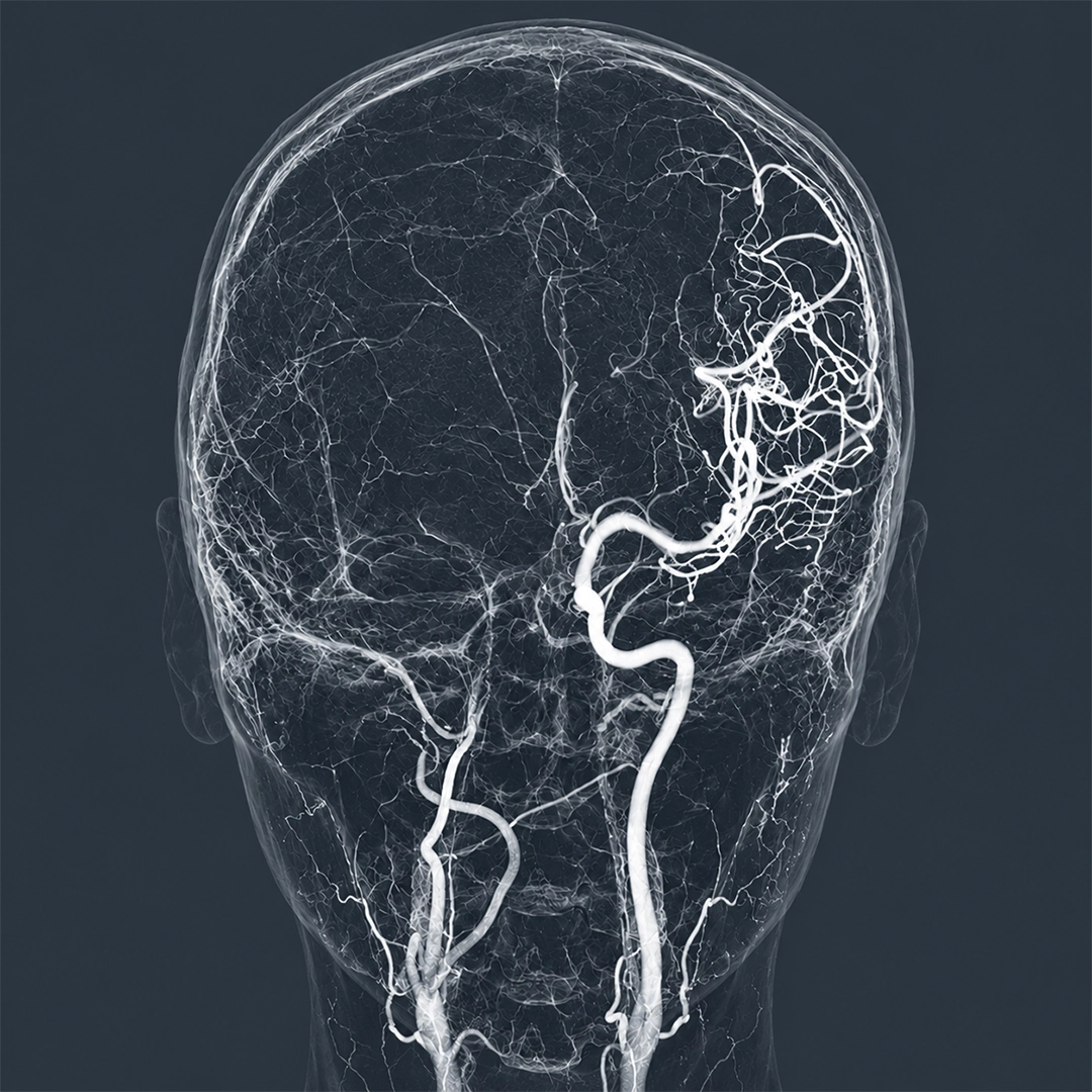
Cerebral Angiogram (DSA) : Brain Blood Vessels Test
A Cerebral Angiogram (Digital Subtraction Angiography – DSA) is an advanced imaging procedure that provides highly detailed images of the brain’s blood vessels. It helps diagnose conditions such as aneurysms, arteriovenous malformations (AVMs), stroke, and other vascular abnormalities, allowing doctors to plan the most appropriate treatment.
What is a Cerebral Angiogram?
A Cerebral Angiogram, also known as Cerebral Angiography or Digital Subtraction Angiography (DSA), is a specialized imaging procedure used to examine the blood vessels of the brain, neck, and spinal cord. It is considered the gold standard for detecting abnormalities in cerebral blood circulation because it provides highly detailed images.
Why is a Cerebral Angiogram Performed?
Cerebral angiogram performed to diagnose or evaluate:
- Brain aneurysms
- Arteriovenous malformations (AVMs)
- Blockage of brain arteries
- Stroke
- Carotid artery disease
- Unexplained brain bleeding
- Brain vascular tumors
- Blood vessel abnormalities before surgery or treatment planning
How is the Procedure Performed?
- The patient lies comfortably on an angiography table.
- Local anaesthesia is administered at the catheter insertion site.
- A small catheter is inserted into an artery in the wrist or groin.
- The catheter is carefully guided to the arteries supplying the brain.
- Contrast dye is injected through the catheter.
- Multiple X-ray images are captured to study blood flow in detail.
- After the procedure, the catheter is removed and pressure is applied to prevent bleeding.
Benefits of Cerebral Angiography
- Provides highly accurate images of brain blood vessels
- Helps diagnose conditions that may not be detected by other imaging tests
- Assists in treatment planning for aneurysms, AVMs, and stroke
- Offers real-time visualization of blood flow
Is Cerebral Angiography Safe?
Yes. Cerebral angiography is generally considered a safe procedure when performed by experienced neurointerventional specialists. Serious complications are uncommon.
Possible Risks and Complications
Although uncommon, potential risks include:
- Bruising or bleeding at the catheter insertion site
- Allergic reaction to contrast dye
- Kidney problems in susceptible patients
- A blood clot forming around the tip of the catheter may rarely lead to neurological complications
Recovery After Cerebral Angiogram
Most patients can return home on the same day or the following day, depending on their condition. Mild soreness or bruising at the catheter site may occur for a few days. Drinking plenty of fluids helps flush the contrast dye from the body. Follow your doctor’s instructions regarding activity restrictions and medications.
Frequently Asked Questions (FAQs)
The procedure is generally not painful. Patients may feel a brief sting from the local anaesthetic and a warm sensation when the contrast dye is injected.
The procedure typically takes approx 30 minutes, depending on the complexity of the examination.
Yes. Most cerebral angiograms are performed under local anaesthesia, and patients remain awake throughout the procedure.
Contrast dye helps highlight blood vessels on X-ray images, allowing doctors to visualize blood flow and identify abnormalities.