Advanced Arteriovenous Malformation (AVM) Diagnosis & Treatment
— Safe, Precise & Minimally Invasive Neurointerventional Care
Brain Arteriovenous Malformation (AVM) – Symptoms, Causes, Diagnosis & Treatment
A Brain Arteriovenous Malformation (AVM) is an abnormal connection between arteries and veins that disrupts normal blood flow and increases the risk of brain hemorrhage, seizures, and neurological complications. Early diagnosis and advanced treatment, can help reduce the risk of bleeding and protect brain function.
What is an Arteriovenous Malformation (AVM)?
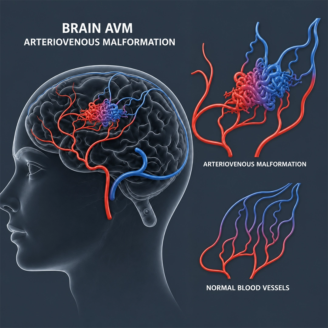
An Arteriovenous Malformation (AVM) is an abnormal tangle of blood vessels where arteries connect directly to veins without the normal network of tiny blood vessels (capillaries) in between. This disrupts normal blood circulation and can place excessive pressure on the blood vessels.
Although AVMs can occur anywhere in the body, Brain AVMs are the most serious because they can lead to brain hemorrhage, seizures, stroke-like symptoms, and other neurological complications.
Many people may have an AVM for years without knowing it, while others develop symptoms that require immediate medical attention.
What Are the Symptoms of an AVM?
The symptoms of a brain AVM vary depending on its size and location. In approximately half of the patients, an AVM is discovered only after it causes bleeding in the brain.
Common AVM symptoms include:
Severe or
recurring headaches
Seizures (fits)
Sudden weakness or numbness in the face, arm, or leg
Difficulty speaking or understanding speech
Vision disturbances
Dizziness or balance problems
Memory or concentration difficulties
Brain hemorrhage (bleeding)
If you experience any sudden neurological symptoms, immediate medical evaluation is essential.
What Is the Risk of Bleeding from an AVM?
- Brain AVMs affect approximately 0.1% of the population. Most AVMs are believed to develop before birth due to abnormal formation of blood vessels.
- The abnormal connection between arteries and veins exposes the veins to high-pressure blood flow, increasing the risk of rupture and bleeding.
Important facts about AVM bleeding risk:
- The annual risk of bleeding is approximately 3–4%.
- AVMs are most likely to bleed between the ages of 10 and 55 years.
- A ruptured AVM can cause a life-threatening brain hemorrhage.
- After an initial bleed, the risk of another hemorrhage significantly increases, especially during the first year.
- The risk remains present until the AVM is successfully treated.
How Is an AVM Diagnosed?
- MRI Brain - MRI is often the first imaging test used to identify a brain AVM. It provides detailed images of the brain and surrounding structures.
- Digital Subtraction Angiography (DSA)- Digital Subtraction Angiography (DSA) is considered the gold standard for AVM diagnosis. It provides highly detailed images of the blood vessels and helps determine:
- The exact size and location of the AVM
- Feeding arteries and draining veins
- Risk of bleeding
- Best treatment approach
In some cases, especially after a recent brain hemorrhage, the AVM may not be clearly visible on MRI or CT scans. DSA helps establish a definitive diagnosis.
AVM Treatment Options
Treatment is individualized based on the size, location, symptoms, and bleeding history of the AVM. The primary goal is to prevent future hemorrhage and protect brain function.
1. Endovascular Embolization
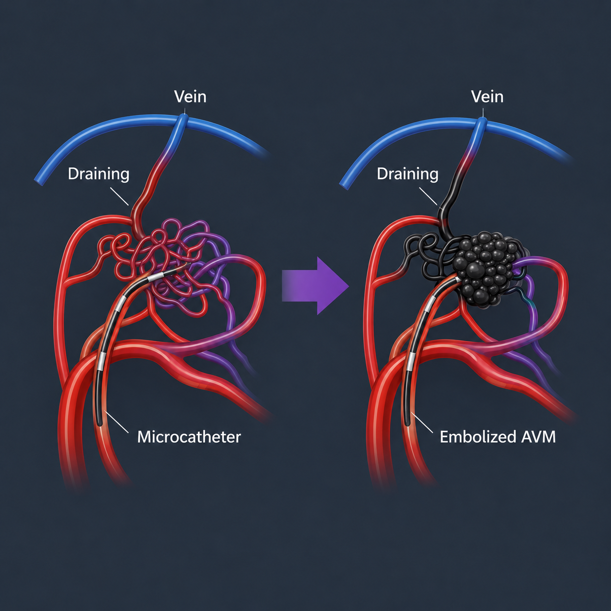
Endovascular Embolization is a minimally invasive procedure performed through a catheter inserted into an artery in the groin.
The catheter is guided to the blood vessels supplying the AVM, and a special embolic material such as Onyx® or medical glue is injected to block blood flow within the abnormal vessels.
Benefits of Embolization
- Minimally invasive treatment
- No large surgical incision
- Reduces blood flow to the AVM
- Can lower the risk of bleeding
- May be used before surgery or radiosurgery
- Faster recovery compared to open surgery
2. Stereotactic Radiotherapy
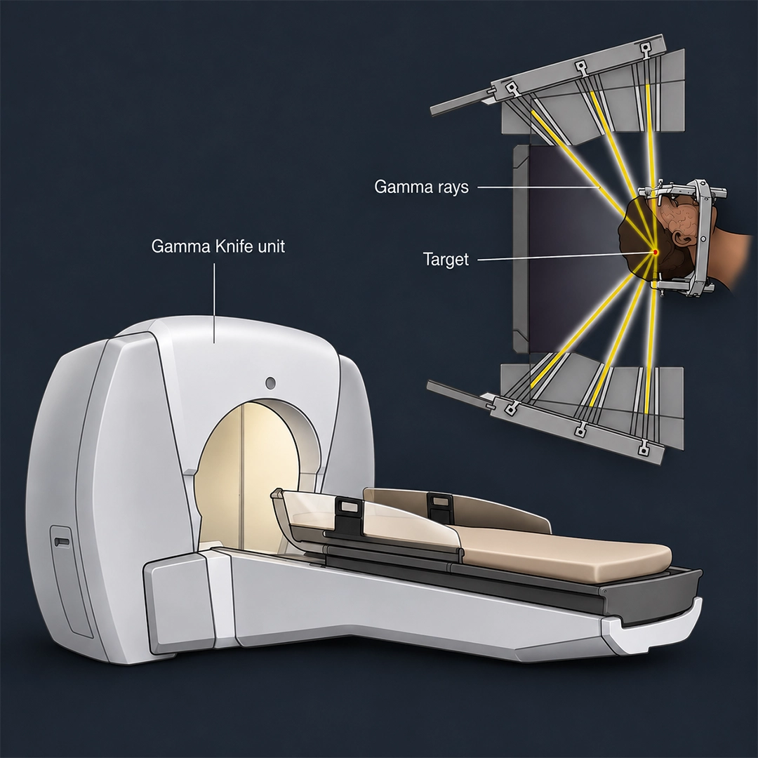
Stereotactic Radiotherapy is a non-invasive treatment that uses highly focused radiation beams to target the AVM.
The radiation gradually causes the abnormal blood vessels to close over time, usually within 3 years in upto 80% of patients.
When Is Radiotherapy Recommended?
- Small AVMs
- Deep-seated AVMs
- AVMs located in critical areas of the brain
- Patients who are not ideal candidates for surgery or embolization.
It is important to note that the risk of bleeding remains until the AVM is completely closed.
3. Microsurgical Excision
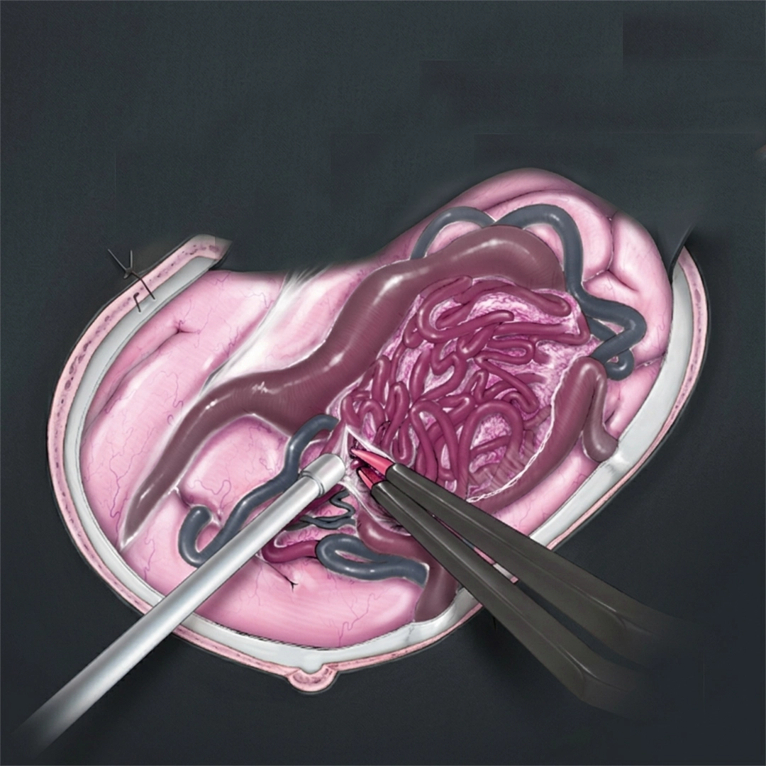
Microsurgical excision is a surgical procedure in which the AVM is removed completely using advanced microsurgical techniques.
This treatment can provide an immediate cure when the entire AVM is successfully removed.
Surgery Is Commonly Recommended When:
- The AVM has already bledSmall AVMs
- A large brain hematoma is present
- The AVM is located near the surface of the brain
- The AVM is situated in a non-critical area of the brain
Deeply located AVMs or those involving important brain functions may not be suitable for surgery due to higher risks.
4. Conservative (Medical) Management
Not every AVM requires immediate intervention.
In selected patients with unruptured AVMs or AVMs that cannot be treated safely, careful observation and regular follow-up may be recommended.
This approach includes:
- Regular MRI or DSA monitoring
- Management of headaches or seizures
- Ongoing neurological evaluation
Although treatment risks are avoided, the AVM remains present and continues to carry a risk of bleeding.
Which AVM Treatment Is Best?
There is no single treatment suitable for every AVM. The best treatment depends on:
Common AVM symptoms include:
AVM size & location
Patient age
Bleeding history
Neurological symptoms
Available expertise
Overall health condition
Frequently Asked Questions (FAQs)
No. An AVM is not a tumor. It is an abnormal connection between arteries and veins.
Yes. An AVM can cause bleeding in the brain, which may result in a hemorrhagic stroke.
Recovery depends on the treatment method used and the complexity of the AVM. Many minimally invasive procedures allow faster recovery than traditional surgery.